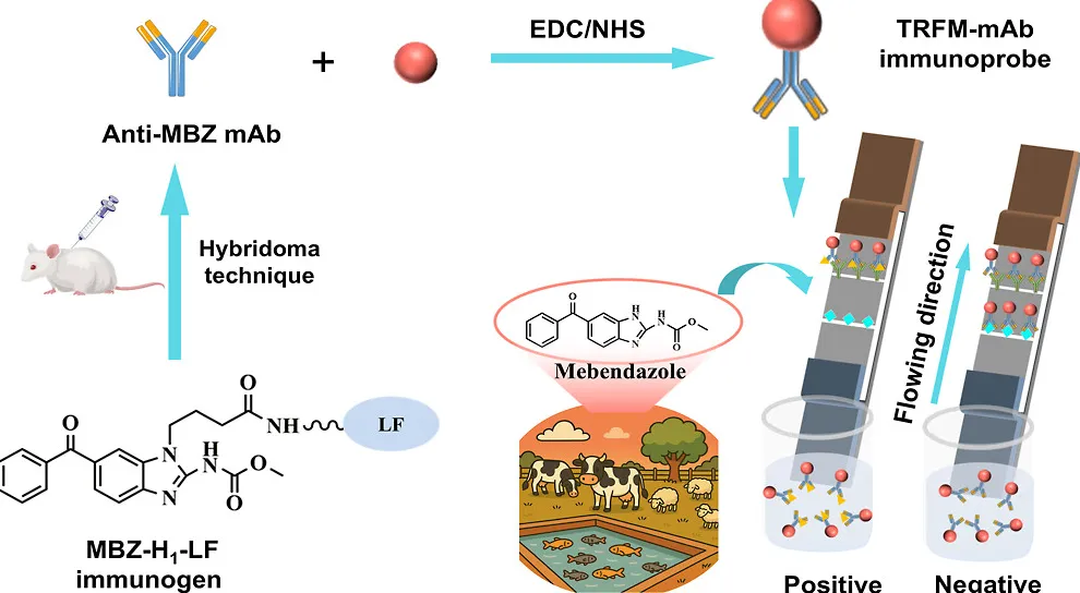
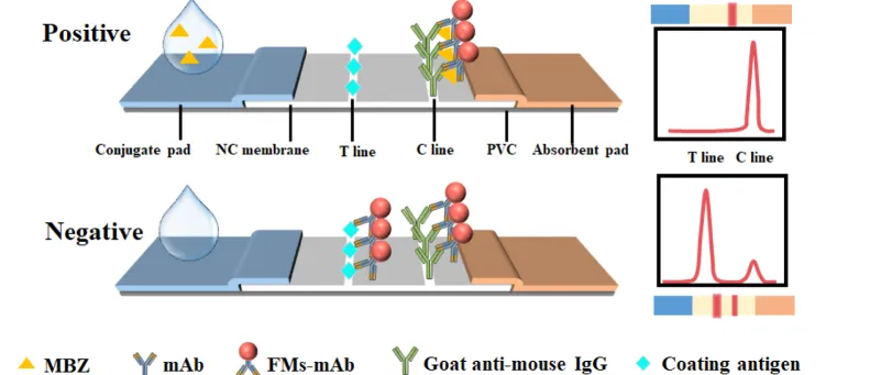
Figure 1. Schematic illustration of TRFIA for MBZ detection.
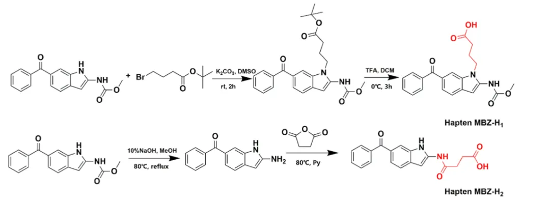
Figure 2. Synthetic route of hapten MBZ-H1 and MBZ-H2.
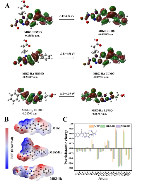
Figure 3. Computational chemistry parameters of MBZ, haptens MBZ-H1 and MBZ-H2 (A); distribution and gaps (ΔE) of the HOMO and LUMO for MBZ, haptens MBZ-H1 and MBZ-H2 (B); ESP surfaces of MBZ, haptens MBZ-H1 and MBZ-H2; (C) Mulliken charge distribution of MBZ, haptens MBZ-H1 and MBZ-H2. Only those atoms that are shared by these structures are displayed. Hydrogen atoms are not included.
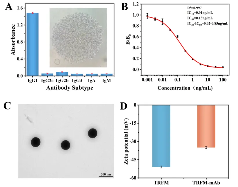
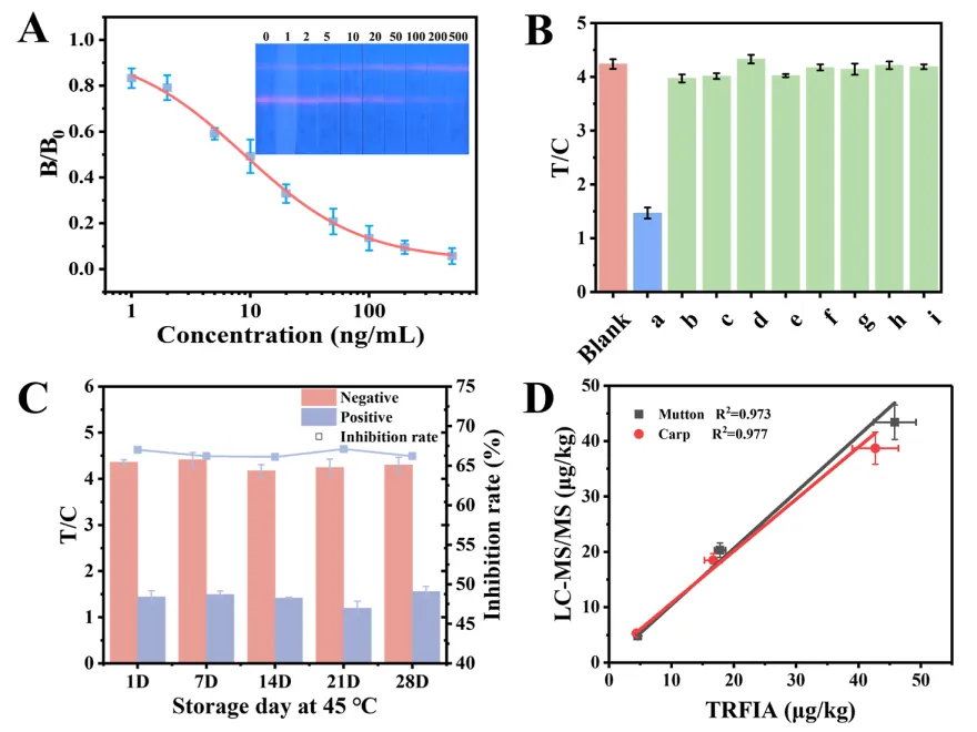
Figure 4. Characterization of the monoclonal antibody (mAb) and TRFM-mAb conjugate. (A) The subtype of the mAb (the inset picture is the prepared hybridoma cell lines under microscopy); (B) the ic-ELISA inhibition curve of the anti-MBZ mAb against MBZ; (C) TEM of TRFM; (D) Zeta potential of TRFM and TRFM-mAb.Figure 5. Performance of TRFIA. (A) The standard curve of MBZ detection by TRFIA, (B) the specificity of TRFIA for detection of MBZ. (a-i: Mebendazole; 2-amino-5-benzoyl-benzimidazole; 5-hydroxymebendazole; Albendazole; Albendazole; Sulfone; Albendazole sulfoxide; Oxfendazole; Flubendazole; Carbendazim); (C) the stability of TRFIA; (D) Comparison the results of LC/MS/MS with those of TRFIA.


 10个月宝宝每天需要喝多少奶粉?
