Chemical Engineering Journal|胃肠道环境中单增李斯特菌通过形成细菌微室增强其对屎肠球菌B1肠溶素A的抗性
近日,南京农业大学食品科技学院叶可萍教授课题组在《Chemical Engineering Journal》发表了题为“Listeria monocytogenes forms bacterial microcompartments to enhance its resistance of enterolysin A in Enterococcus faecium B1 under gastrointestinal conditions”的研究论文(一区,IF=13.2)。该研究围绕屎肠球菌B1在胃肠道条件下对单增李斯特菌的拮抗机制及病原菌的适应性应答展开,发现屎肠球菌B1不仅可显著抑制单增李斯特菌在体外胃肠道消化过程中的存活及毒力表达,还可减轻小鼠感染水平。研究进一步证实,单增李斯特菌在屎肠球菌B1胁迫下会形成细菌微室以增强环境耐受,而屎肠球菌B1中的enlA基因是介导这一过程并发挥抑菌作用的关键因素。该研究揭示了乳酸菌与食源性病原菌在胃肠环境中的互作新机制,为发酵食品安全控制及益生菌资源开发提供了新的理论依据。
单增李斯特菌是重要的食源性致病菌之一,能够耐受冷藏、酸性和胆盐等多种不利环境,常可随受污染食品进入人体消化道,并在通过胃肠道屏障后进一步引发系统性感染,因此其在食品安全与人体健康领域一直备受关注。乳酸菌作为食品和肠道环境中的重要有益微生物,被认为在抑制食源性病原菌方面具有较大应用潜力,其中屎肠球菌因能够产生细菌素、参与营养竞争并占据黏附位点而显示出较强的拮抗作用。然而,在胃肠道条件下,屎肠球菌B1抑制单增李斯特菌的作用机制,以及单增李斯特菌对屎肠球菌B1拮抗作用的适应性应答机制,仍有待进一步阐明。基于此,本研究首先通过小鼠感染模型评价屎肠球菌B1对单增李斯特菌体内感染的缓解作用,随后结合体外模拟胃肠道消化模型,从病原菌存活能力、毒力特征和应激响应等层面解析两者在消化环境中的互作规律,并进一步利用转录组学、基因缺失与互补验证以及透射电镜观察等手段,重点揭示屎肠球菌B1中enlA基因及单增李斯特菌细菌微室形成在这一互作过程中的作用,为认识乳酸菌与食源性病原菌在胃肠环境中的互作机制提供了新的研究视角。
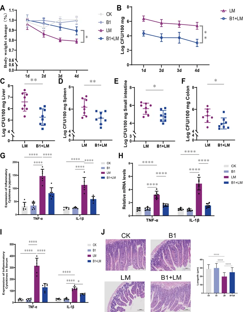
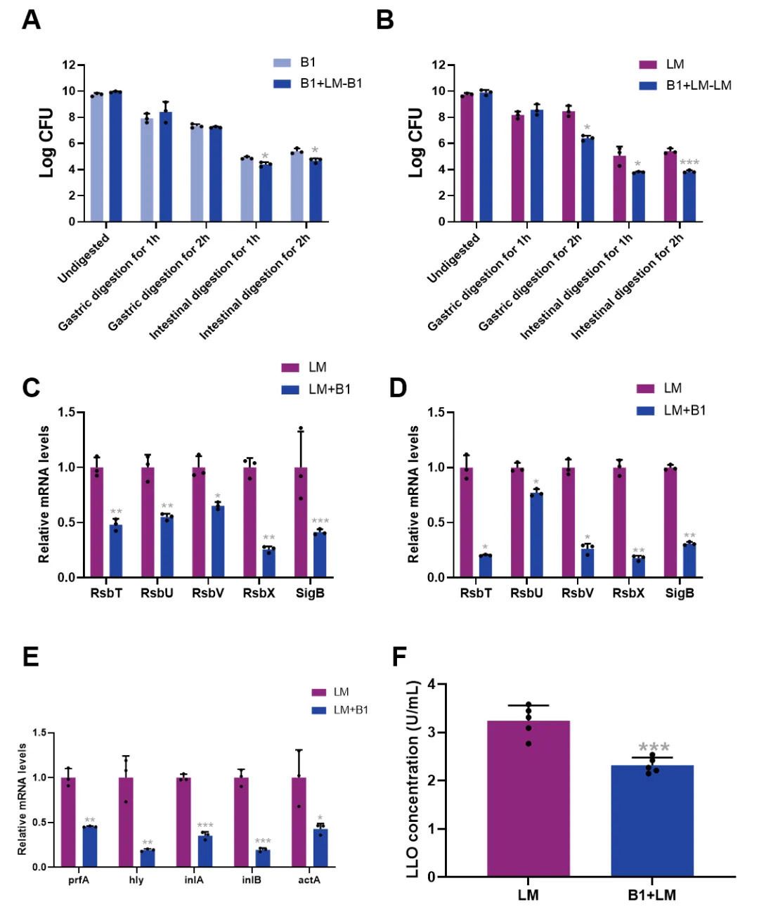
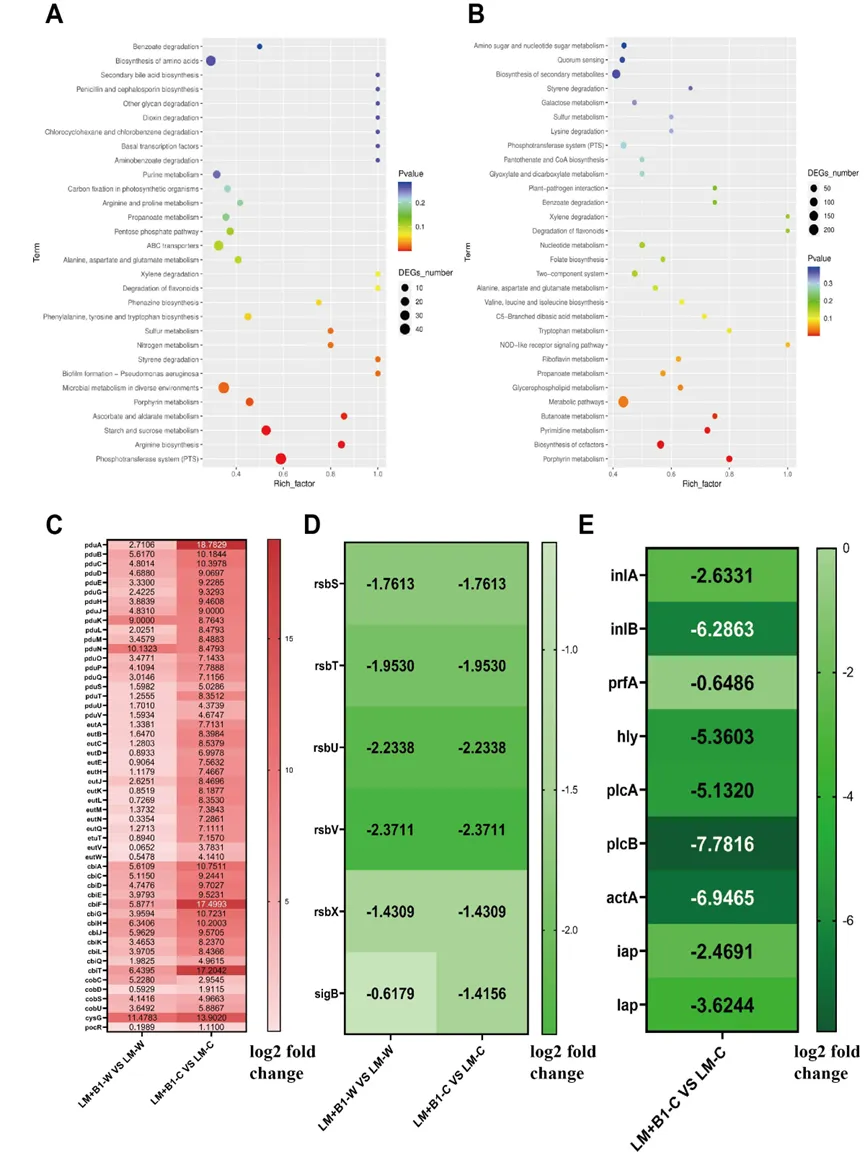
屎肠球菌B1可减轻小鼠单增李斯特菌感染引起的体重下降、降低肝脏、脾脏及肠道组织中的菌载量,并缓解空肠炎症反应。进一步的,在体外模拟胃肠道消化条件下,屎肠球菌B1可显著抑制单增李斯特菌存活,降低其sigB相关应激响应及多种毒力基因表达。值得注意的是,单增李斯特菌在屎肠球菌B1拮抗压力下会形成细菌微室,以增强其在胃肠道环境中的适应能力。同时,屎肠球菌B1中的enlA基因在抑制单增李斯特菌及诱导其细菌微室形成过程中发挥关键作用。该研究揭示了乳酸菌与食源性病原菌在胃肠环境中的互作新机制,为发酵食品安全控制及功能菌株开发提供了新的理论依据。
Figure 1
Figure 1 E. faecium B1 alleviated L. monocytogenes infection in mice. (A) The body weight change of mice after L. monocytogenes infection, n=8; (B-F) The L. monocytogenes loads in feces, liver, spleen, small intestine, and colon, respectively; (G) The concentrations of the pro-inflammatory cytokines TNF-α and IL-1β in jejunum of mice; (H) Relative mRNA levels of inflammatory response-related genes TNF-α and IL-1β in jejunum of mice; (I) The concentrations of the pro-inflammatory cytokines TNF-α and IL-1β in serum of mice; (J) HE staine of jejunum in mice. Significance levels are shown as follows: *P < 0.05, **P < 0.01, ***P < 0.001, and ****P < 0.0001.
Figure 2
Figure 2 The effects of E. faecium B1 on the states of L. monocytogenes during in vitro gastrointestinal digestion. (A) Survival counts of E. faecium B1 in the B1+LM and B1 group; (B) Survival counts of L. monocytogenes in the B1+LM and LM group; (C-D) The expression of sigB-related genes in L. monocytogenes after gastric and intestinal digestion, respectively; (E-F) The expression of virulence genes and LLO protein in L. monocytogenes after gastrointestinal digestion; Significance levels are shown as follows: *P < 0.05, **P < 0.01, ***P < 0.001, and ****P < 0.0001.
Figure 3
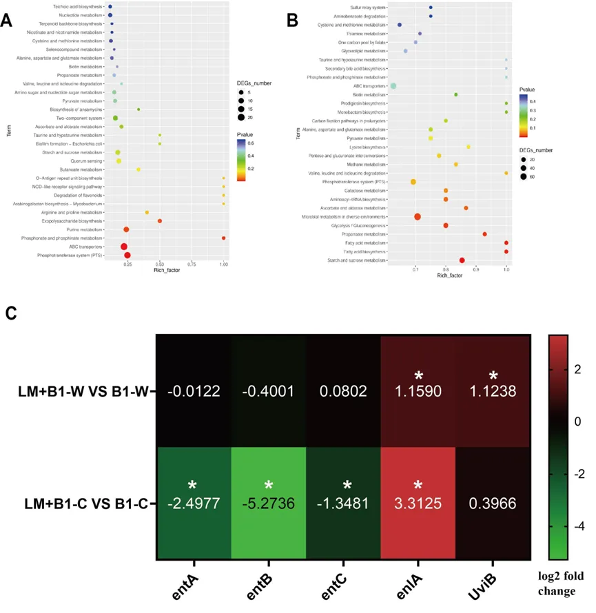
Figure 3 The transcriptomic investigation of L. monocytogenes during in vitro gastrointestinal digestion. (A-B) KEGG pathway analysis in L. monocytogenes after gastric and intestinal digestion, respectively (comparison of B1+LM vs LM); (C-D) The expression of genes related to bacterial microcompartment and sigB signaling pathway in L. monocytogenes after gastric digestion and intestinal digestion, respectively (comparison of B1+LM vs LM); (E)The expression of genes related to virulence in L. monocytogenes after intestinal digestion (comparison of B1+LM vs LM). LM-W and LM-C indicate the transcriptomic analysis of L. monocytogenes after gastric digestion and intestinal digestion, respectively; LM+B1-W and LM+B1-C indicate the transcriptomic analysis of L. monocytogenes in the LM+B1 group after gastric digestion and intestinal digestion, respectively. Genes shown in the heatmaps met the significance threshold of P < 0.05 in transcriptomic analysis.
Figure 4
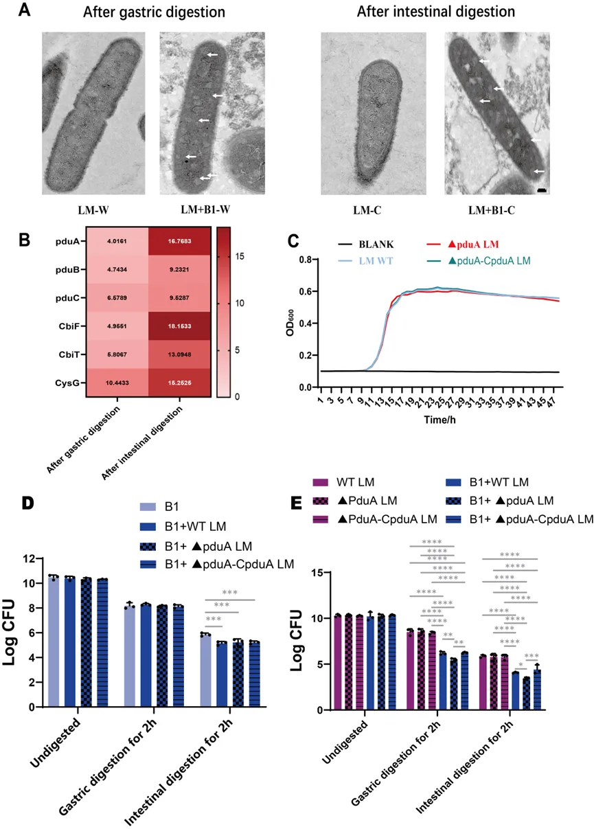
Figure 4 Role of the pduA gene in modulating L. monocytogenes resistance to E. faecium B1 during in vitro gastrointestinal digestion. (A) Transmission electron microscopy observation of L. monocytogenes, the scale bar was 100 nm; (B) The expression of bacterial microcompartment-related genes (comparison of B1+LM vs LM), genes shown in the heatmaps met the significance threshold of P < 0.05; (C) Growth curve of L. monocytogenes; (D) Survival counts of E. faecium B1 in the B1+LM and B1 group; (E) Survival counts of L. monocytogenes in the B1+LM and LM group. Significance levels are shown as follows: *P < 0.05, **P < 0.01, ***P < 0.001, and ****P < 0.0001.
Figure 5
Figure 5 The transcriptomic investigation of E. faecium B1 during in vitro gastrointestinal digestion. (A-B) KEGG pathway analysis in E. faecium after gastric and intestinal digestion, respectively (comparison of B1+LM vs B1); (C) The expression of genes related to bacteriocin in E. faecium after gastric and intestinal digestion, respectively (comparison of B1+LM vs B1). B1-W and B1-C indicate the transcriptomic analysis of E. faecium B1 after gastric digestion and intestinal digestion, respectively; LM+B1-W and LM+B1-C indicate the transcriptomic analysis of E. faecium B1 in the LM+B1 group after gastric digestion and intestinal digestion, respectively.
Figure 6
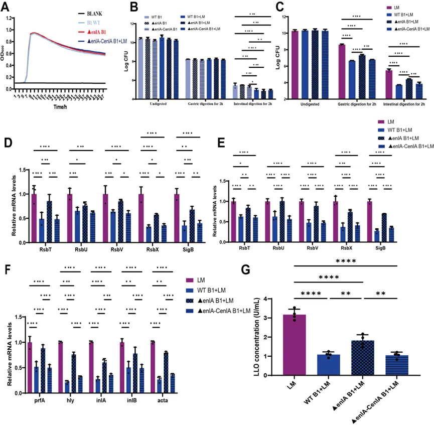
Figure 6 Modulation of L. monocytogenes survival by the enlA gene of E. faecium B1 during in vitro gastrointestinal digestion. (A) Growth curve of E. faecium; (B) Survival counts of E. faecium; (C) Survival counts of L. monocytogenes; (D-E) The expression of sigB-related genes in L. monocytogenes after gastric and intestinal digestion, respectively. (F-G) The expression of virulence genes and LLO protein in L. monocytogenes after gastrointestinal digestion. Significance levels are shown as follows: *P < 0.05, **P < 0.01, ***P < 0.001, and ****P < 0.0001.
Figure 7
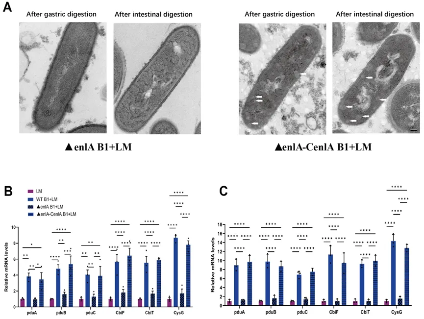
Figure 7 The effect of E. faecium enlA gene expression on L. monocytogenes bacterial microcompartment formation. (A) Transmission electron microscopy observation of L. monocytogenes, the scale bar was 100 nm;(B-C) The expression of bacterial microcompartment-related genes in L. monocytogenes after gastric and intestinal digestion, respectively. Significance levels are shown as follows: *P < 0.05, **P < 0.01, ***P < 0.001, and ****P < 0.0001.
Figure 8
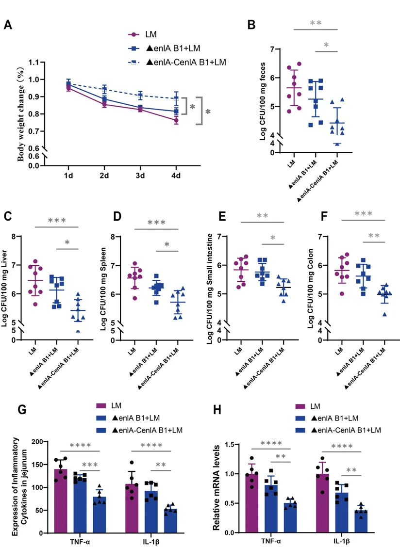
Figure 8 E. faecium B1 enlA gene-mediated inhibition of L. monocytogenes infection in mice. (A) The body weight change of mice after L. monocytogenes infection, n=8; (B-F) The L. monocytogenes loads in feces, liver, spleen, small intestine, and colon, respectively; (G) The concentrations of the pro-inflammatory cytokines TNF-α and IL-1β in the jejunum of mice; (H) Relative mRNA levels of inflammatory response-related genes TNF-α and IL-1β in the jejunum of mice. Significance levels are shown as follows: *P < 0.05, **P < 0.01, ***P < 0.001, and ****P < 0.0001.
叶可萍
南京农业大学教授、博士生导师,现任国家肉品质量安全控制工程技术研究中心副主任,南京农业大学食品科技学院食品质量与安全系主任,农业农村部肉及肉制品质量检验测试中心(南京)质量负责人。近年来主要围绕肉品品质及贮藏保鲜、肉品中病原菌预测与控制、真菌蛋白仿制肉等方向开展研究工作。主持国家自然科学基金面上项目和青年项目3项,国家“十四五”重点研发计划项目课题、国家“十四五”重点研发计划揭榜挂帅项目任务,以及江苏省自然科学基金面上项目、新疆重大科技专项任务、国家食品安全风险评估中心项目和企业重大产学研合作项目等20余项。以第一作者或通信作者在Chemical Engineering Journal、Food Hydrocolloids、Food Chemistry、Food Control、Food Packaging and Shelf Life、Frontiers in Cellular and Infection Microbiology、International Journal of Molecular Sciences、Frontiers in Microbiology、Meat Science、Food Research International等期刊发表论文70余篇。申请发明专利5项,获授权发明专利2项、实用新型专利2项,参与制定标准1项,参编教材与书籍2部,获中国食品科学技术学会科技创新一等奖。
https://doi.org/10.1016/j.cej.2026.175904
免责声明:「原创」仅代表原创编译,水平有限,仅供学术交流,本平台不主张原文的版权,如有侵权,请联系删除。文献解读或作者简历如有疏漏之处,我们深表歉意,请作者团队及时联系《食探未来》主编(微信号:shitanweilai8077),我们会在第一时间进行修改或撤稿重发,感谢您的谅解!


 10个月宝宝每天需要喝多少奶粉?
